
Automated Colon Disease Classification via Wireless Capsule Endoscopy & YOLOv8
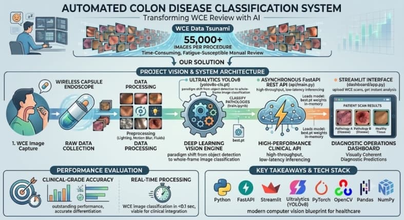
Wireless Capsule Endoscopy (WCE) has revolutionized gastroenterology by providing a non-invasive method to examine the entire gastrointestinal tract. However, a single WCE procedure generates tens of thousands of images. For a gastroenterologist, manually reviewing this massive volume of data to find microscopic signs of disease is incredibly time-consuming and highly susceptible to human fatigue.
This Deep Dive introduces our Automated Colon Disease Classification System. By deploying state-of-the-art computer vision algorithms—specifically YOLOv8—this pipeline autonomously processes WCE frames to identify and classify colon anomalies, transforming a tedious manual review process into a rapid, data-driven diagnostic workflow.
Project Vision
Our objective was to architect a comprehensive clinical tool rather than a standalone script. The vision encompassed training an ultra-fast vision model capable of distinguishing complex gastrointestinal pathologies, wrapping it within a high-throughput REST API, and providing a user-friendly dashboard where medical professionals can upload scans for instant analysis.
Technical Hurdles
Complex Biological Imagery: Endoscopic images are notoriously difficult for AI due to extreme variations in lighting, motion blur, and obscuring biological fluids. Robust data preprocessing and augmentation were mandatory to help the model extract meaningful features.
High-Throughput Requirements: Since clinical application requires processing thousands of frames per patient in a reasonable timeframe, the underlying neural network had to be aggressively optimized for inference speed without sacrificing diagnostic precision.
System Architecture
The architecture was designed for both high performance and modularity, consisting of three main engineering pillars:
1 Deep Learning Vision Engine
YOLOv8 Classification (train.ipynb): We utilized the Ultralytics YOLOv8 architecture (yolov8s-cls.pt), shifting its paradigm from typical object detection to high-speed image classification. This allowed the system to quickly categorize whole frames into specific disease states.
2 High-Performance Clinical API
FastAPI Backend (api/main.py): To make the model accessible, we engineered an asynchronous REST API. This service loads the optimized training weights (best.pt) into memory and handles incoming image inference requests with minimal latency.
3 Diagnostic Operations Dashboard
Interactive Interface (dashboard/app.py): A dedicated Streamlit dashboard was built for the end-user (clinicians). It securely communicates with the API, allowing users to upload WCE imagery and receive immediate, visually coherent diagnostic predictions.
Performance Evaluation
Clinical-Grade Accuracy: The YOLOv8 model demonstrated outstanding metrics across validation datasets, accurately differentiating between various colon diseases and healthy tissue.
Real-Time Processing: By decoupling the heavy inference engine into a dedicated API, the dashboard processes and classifies WCE images in a fraction of a second, proving its viability for live clinical integration.
Key Takeaways & Tech Stack
This project illustrates the immense potential of deploying modern computer vision architectures in healthcare. By optimizing YOLOv8 for classification and serving it through a scalable backend, we provided a blueprint for how AI can alleviate the diagnostic burden on medical professionals.
Languages & Frameworks: Python, FastAPI, Streamlit.
Computer Vision: Ultralytics (YOLOv8), PyTorch, OpenCV.
Data Processing: Pandas, NumPy.